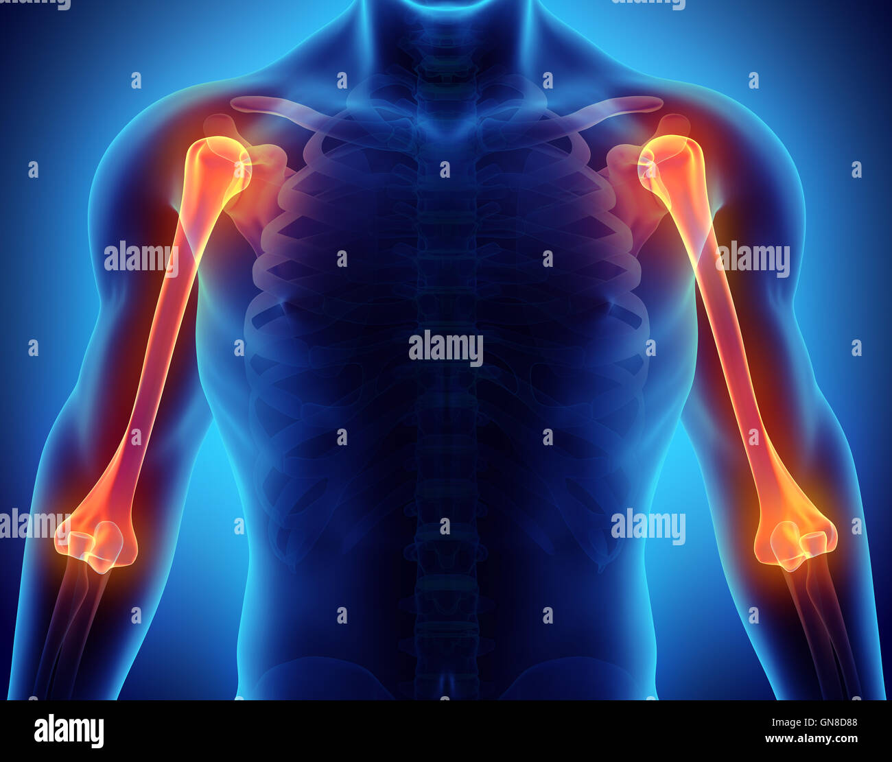
The human humerus is the longest and largest bone of the upper extremity, extending from the shoulder joint to the elbow joint. It serves as a critical structural pillar for the arm, providing an attachment site for numerous muscles that control shoulder and elbow mobility while protecting vital neurovascular pathways.
Introduction to the Human Humerus
The humerus is a typical long bone characterized by a central shaft (diaphysis) and two distinct extremities (epiphyses), known as the proximal and distal ends. It functions primarily as a mechanical lever for the upper limb, enabling a wide range of motion from heavy lifting to fine motor positioning. Because it bridges the torso to the forearm, the humerus must endure significant biomechanical forces, including compression, tension, torsion, and bending.
Understanding the anatomy of the humerus requires examining its specialized bony landmarks, such as the head, tubercles, epicondyles, and fossae. These features not only facilitate joint articulation with the scapula (shoulder blade) and the radius and ulna (forearm bones), but they also anchor the rotator cuff and major arm muscles. Clinically, the humerus is vulnerable to high-energy trauma, pathological fractures due to bone diseases, and nerve compression syndromes. Recognizing its structural organization is essential for diagnosing injuries, planning surgical interventions, and managing rehabilitation effectively.
(Note: The following anatomical and clinical overview is provided for informational and educational purposes. Individuals seeking medical evaluation for an injury or condition related to the upper arm should consult a qualified healthcare professional.)
Proximal Anatomy of the Humerus
The proximal extremity of the humerus articulates with the glenoid cavity of the scapula to form the glenohumeral (shoulder) joint, a ball-and-socket articulation characterized by high mobility and inherent structural laxity.
The Head of the Humerus and Neck Regions
The most prominent feature of the proximal humerus is the humeral head, a smooth, hemispherical articular surface directed medially, upward, and slightly backward. It is covered in hyaline cartilage and fits into the shallow glenoid fossa.
Immediately surrounding the articular margin of the head is the anatomical neck, a slight circumferential constriction separating the head from the greater and lesser tubercles. In contrast, the surgical neck lies distal to the tubercles, marking the transition from the rounded proximal epiphysis to the cylindrical shaft. The surgical neck is clinically significant because it is a frequent site of osteoporotic and traumatic fractures.
Greater and Lesser Tubercles
Projecting laterally from the anatomical neck is the greater tubercle, which presents three flattened impressions for the insertion of rotator cuff muscles:
- Superior facet: Insertion site for the supraspinatus muscle.
- Middle facet: Insertion site for the infraspinatus muscle.
- Inferior facet: Insertion site for the teres minor muscle.
The lesser tubercle is smaller and projects anteriorly, serving as the primary insertion point for the subscapularis muscle.
Separating these two tubercles is a deep groove known as the intertubercular sulcus (or bicipital groove). This channel lodges the tendon of the long head of the biceps brachii muscle as it ascends toward the supraglenoid tubercle of the scapula. The groove features medial and lateral lips, which serve as attachment sites for the pectoralis major, latissimus dorsi, and teres major muscles.
Shaft of the Humerus (Diaphysis)
The shaft of the humerus transitions from a cylindrical shape proximally to a triangular, flattened shape distally. It features three distinct borders and three surfaces, providing extensive surface area for muscular attachment.
Borders and Surfaces
- Anterior border: Runs downward from the anterior margin of the greater tubercle, dividing inferiorly to enclose the coronoid fossa.
- Medial border: Extends from the lesser tubercle to the medial epicondyle, featuring a rough ridge for the coracobrachialis muscle insertion.
- Lateral border: Extends from the greater tubercle to the lateral epicondyle, interrupted midway by the spiral groove.
The surfaces—anteromedial, anterolateral, and posterior—accommodate large muscle groups. The deltoid tuberosity is a V-shaped roughened area located on the anterolateral surface of the mid-shaft where the deltoid muscle inserts. On the posterior surface, running obliquely from medial to lateral, is the radial groove (or spiral groove). This channel transmits the radial nerve and the profunda brachii artery as they wrap around the bone.
| Region / Landmark | Primary Anatomical Feature | Associated Muscles or Structures |
|---|---|---|
| Proximal End | Humeral Head & Tubercles | Rotator cuff (supraspinatus, infraspinatus, teres minor, subscapularis) |
| Bicipital Groove | Intertubercular Sulcus | Tendon of the long head of the biceps brachii |
| Shaft (Mid-Diaphysis) | Deltoid Tuberosity | Insertion of the deltoid muscle |
| Radial Groove | Posterior Spiral Depression | Radial nerve and profunda brachii artery |
| Distal End | Trochlea and Capitulum | Articulation with ulna and radius; flexor/extensor origins |
Distal Anatomy and Articulations
The distal extremity of the humerus widens transversely, forming a flattened condyle that articulates with the forearm bones to establish the elbow and radioulnar mechanics.
Articular Surfaces: Trochlea and Capitulum
The distal articular surface is divided into two primary regions:
- Trochlea: A spool-shaped surface located medially that articulates with the trochlear notch of the ulna. It allows primarily for hinge-like flexion and extension of the elbow.
- Capitulum: A rounded, smooth eminence located laterally that articulates with the concave superior surface of the radial head, facilitating both elbow flexion and forearm rotation (pronation and supination).
Non-Articular Landmarks: Epicondyles and Fossae
Flanking the articular surfaces are two prominent bony projections:
- Medial epicondyle: Larger and more prominent than its lateral counterpart. It serves as the common origin for the flexor muscles of the forearm and protects the ulnar nerve as it passes posteriorly through the ulnar sulcus (the "funny bone" area).
- Lateral epicondyle: Serves as the common origin for the extensor muscles of the forearm.
Above the articular surfaces, three depressions accommodate the forearm bones during extreme ranges of motion:
- Radial fossa: Located anteriorly above the capitulum, accommodating the head of the radius during full elbow flexion.
- Coronoid fossa: Located anteriorly above the trochlea, receiving the coronoid process of the ulna during flexion.
- Olecranon fossa: A deep depression located posteriorly above the trochlea that houses the olecranon process of the ulna during full elbow extension.
Biomechanical Role and Muscle Attachments
The human humerus acts as a class-three lever system in most upper limb movements, where the effort force is applied between the fulcrum (shoulder or elbow joint) and the load. This mechanical arrangement prioritizes speed and range of motion over raw lifting force.
Numerous muscles across the shoulder girdle, thorax, and arm anchor directly to the humerus:
- Shoulder Abduction: Driven primarily by the deltoid, which inserts at the deltoid tuberosity.
- Internal Rotation: Mediated by the subscapularis, pectoralis major, latissimus dorsi, and teres major.
- External Rotation: Powered by the infraspinatus and teres minor.
- Flexion and Extension: Coordinated by the biceps brachii, brachialis, coracobrachialis, and triceps brachii.
This intricate network requires precise load distribution. Cartilage preservation on the humeral head and smooth gliding within the bicipital and radial grooves are critical for maintaining fluid arm kinematics.
Clinical Conditions and Common Injuries
Because of its length, position, and exposure to physical stress, the humerus is susceptible to several distinct clinical conditions and traumatic injuries.
Fractures of the Humerus
Humerus fractures are generally classified by their anatomical location:
- Proximal Humerus Fractures: Common in elderly individuals with osteoporosis following a fall onto an outstretched hand (FOOSH). They may be nondisplaced or severely comminuted, often managed conservatively with a sling or surgically with plates, screws, or shoulder hemiarthroplasty.
- Humerus Shaft Fractures: Typically result from high-energy direct trauma (such as motor vehicle collisions) or torsional forces (such as arm wrestling). Because the radial nerve runs in close contact with the middle and distal thirds of the radial groove, shaft fractures carry a notable risk of radial nerve palsy, leading to wrist drop.
- Distal Humerus Fractures: Include supracondylar fractures (frequent in pediatric populations) and intercondylar T- or Y-shaped fractures in adults. These often require open reduction and internal fixation (ORIF) to restore joint congruity and prevent post-traumatic arthritis or joint contractures.
Nerve and Vascular Vulnerabilities
The close proximity of major neurovascular structures to the humeral shaft makes specific fracture patterns hazardous:
- Axillary Nerve: Vulnerable to proximal fractures or anterior shoulder dislocations occurring near the surgical neck.
- Radial Nerve: At risk in mid-shaft spiral fractures. Injury results in the inability to extend the wrist and fingers.
- Ulnar Nerve: Traversed near the medial epicondyle; chronic irritation or acute trauma can lead to paresthesia or weakness in the intrinsic hand muscles.
Frequently Asked Questions
What type of bone is the human humerus?
The human humerus is classified as a long bone. It features a tubular shaft (diaphysis) composed of compact cortical bone enclosing a medullary cavity, and two expanded articular ends (epiphysis) containing spongy trabecular bone covered by a thin layer of compact bone and articular cartilage.
Which nerves are most at risk during a humerus fracture?
The radial nerve is most commonly injured during fractures of the mid-shaft of the humerus due to its close anatomical path along the radial groove. The axillary nerve is frequently at risk during proximal fractures near the surgical neck, and the ulnar nerve can be affected by distal injuries near the medial epicondyle.
What is the difference between the anatomical neck and the surgical neck?
The anatomical neck is a slight groove immediately adjacent to the articular surface of the humeral head, separating the head from the tubercles. The surgical neck is located more distally, just below the greater and lesser tubercles, where the cylindrical shaft begins. The surgical neck is clinically significant because it is a frequent site of fractures.
How does the humerus contribute to elbow movement?
The distal end of the humerus features two distinct articular surfaces that form the elbow joint. The trochlea articulates with the ulna to create a hinge joint allowing flexion and extension, while the capitulum articulates with the radius to support both flexion and rotational movements of the forearm.
Can a human humerus heal without surgery?
Many humerus fractures—particularly nondisplaced proximal fractures or stable shaft fractures—heal successfully without surgery through immobilization using casts, functional braces, or slings. However, displaced fractures, intra-articular fractures involving the elbow, or fractures complicated by nerve entrapment typically require surgical intervention.
Conclusion and Next Actions
The human humerus is a marvel of skeletal engineering, providing structural integrity, extensive muscular anchorage, and protection for vital neurovascular pathways. Recognizing its proximal, diaphyseal, and distal anatomy clarifies how the upper limb achieves its remarkable dexterity and strength. For individuals managing upper arm trauma, persistent shoulder or elbow pain, or suspected fractures, prompt medical evaluation by an orthopedic specialist is essential to ensure accurate diagnosis, appropriate stabilization, and full functional recovery.