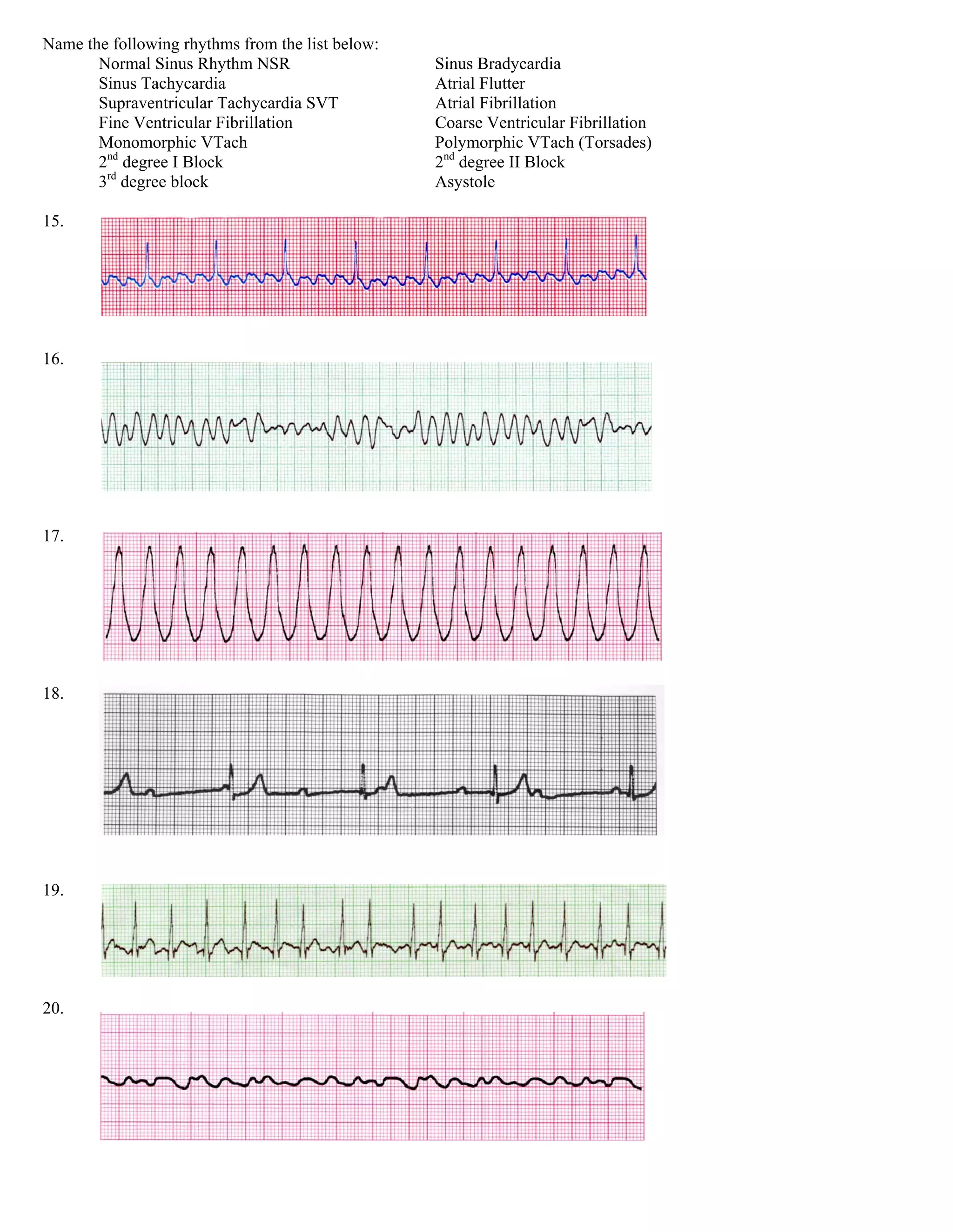
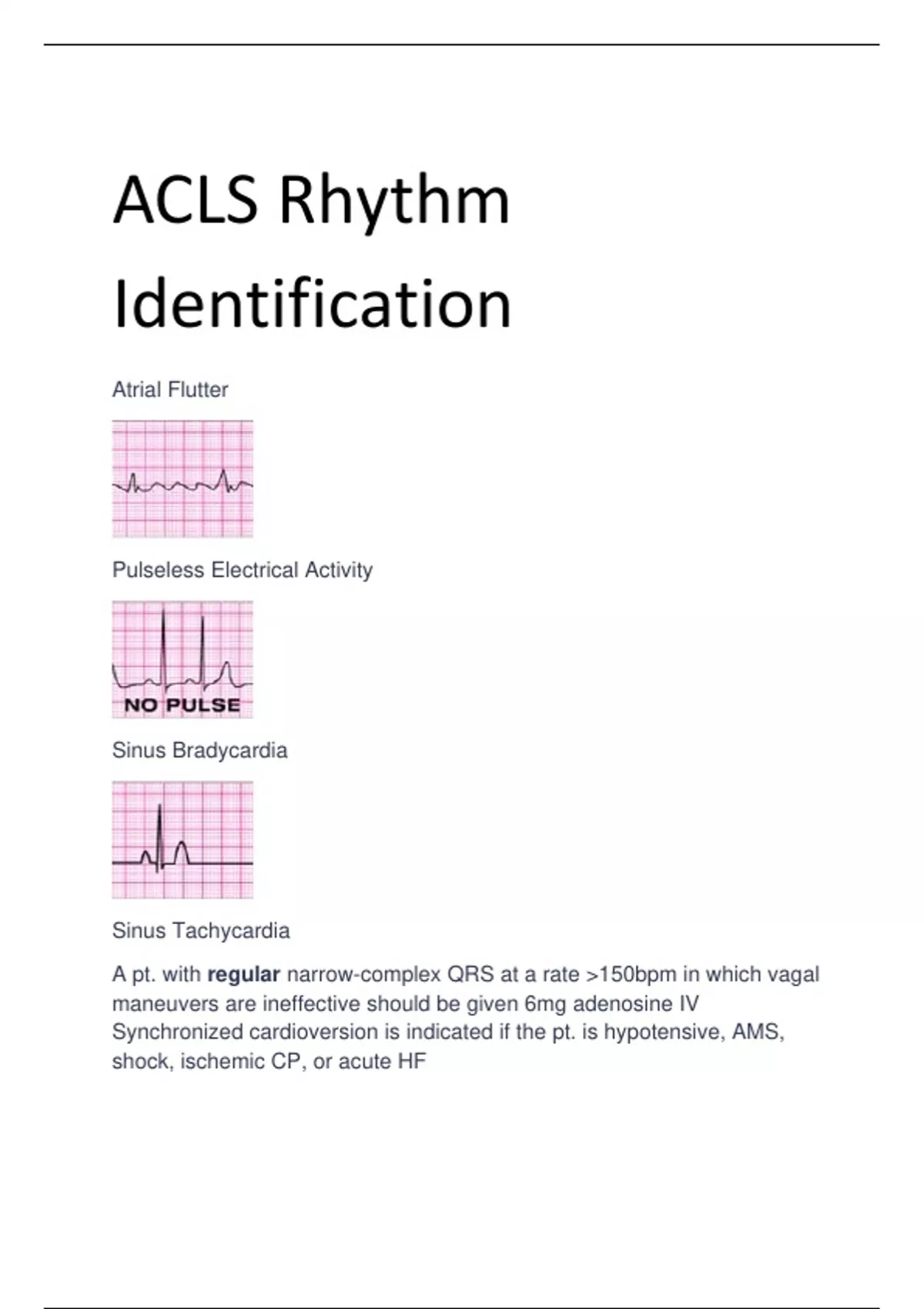
Electrocardiogram (ECG) rhythm recognition in Advanced Cardiovascular Life Support (ACLS) forms the foundation of rapid cardiac emergency management. ACLS rhythms are categorized into three major clinical presentations: cardiac arrest (shockable versus non-shockable), symptomatic bradycardia, and stable or unstable tachycardia. Successful patient management requires systematic ECG analysis coupled with accurate adherence to standardized American Heart Association (AHA) algorithms.
Disclaimer: This article serves as an educational reference for healthcare professionals reviewing cardiac electrophysiology and standardized resuscitation guidelines. It does not replace formal ACLS certification, clinical judgment, or institutional emergency protocols.
Foundations of Systematic ECG Interpretation in ACLS
In an emergency setting, evaluating a cardiac rhythm under ACLS guidelines requires a rapid, reproducible framework. Rather than memorizing static rhythm strips, clinicians analyze continuous cardiac telemetry or a 12-lead ECG using five fundamental parameters.
1. Heart Rate Evaluation
Determine whether the intrinsic ventricular rate is slow (<60 beats per minute), normal (60–100 bpm), or fast (>100 bpm). In severe tachyarrhythmias, rates frequently exceed 150 bpm, which is a key clinical threshold in the ACLS Tachycardia Algorithm for distinguishing primary arrhythmias from compensatory sinus tachycardia.
2. Rhythm Regularity
Examine the R-R intervals across the rhythm strip. Regular rhythms display uniform spacing between consecutive R waves, whereas irregular rhythms exhibit variable R-R intervals. Irregularity can be regularly irregular (such as recurrent Mobitz Type I AV block patterns) or irregularly irregular (characteristic of atrial fibrillation).
3. P Wave Morphology and Presence
Identify whether distinct P waves precede every QRS complex. Upright, uniform P waves in lead II signal a sinus origin. Absent, inverted, fibrillatory, or flutter waves point toward ectopic or reentrant mechanisms within the atria.
4. PR Interval Measurement
Measure the time from the onset of the P wave to the beginning of the QRS complex. The normal PR interval ranges from 0.12 to 0.20 seconds (3 to 5 small boxes). Prolonged PR intervals suggest conduction delays through the atrioventricular (AV) node, whereas progressive PR lengthening indicates specific heart block subtypes.
5. QRS Complex Duration and Architecture
Measure the width of the QRS complex from its initial deflection to the J-point. A narrow QRS duration (<0.12 seconds) indicates normal, rapid ventricular depolarization via the His-Purkinje system. A wide QRS duration (≥0.12 seconds) signifies delayed intraventricular conduction, originating either from ventricular foci (e.g., ventricular tachycardia) or aberrant supraventricular conduction (e.g., bundle branch block).
Cardiac Arrest Rhythms (The Cardiac Arrest Algorithm)
Cardiac arrest algorithms divide rhythms into two functional groups: shockable rhythms, where immediate electrical defibrillation is indicated, and non-shockable rhythms, which rely on high-quality cardiopulmonary resuscitation (CPR) and prompt administration of intravenous or intraosseous epinephrine.
- Patient in Cardiac Arrest?
- Evaluate Rhythm via Monitor:
- Shockable Rhythms
- Ventricular Fibrillation (VF)
- Pulseless Ventricular Tachycardia (pVT)
- Interventions:
- Defibrillate
- Continuous CPR
- Epinephrine/Amiodarone
- Non-Shockable Rhythms
- Asystole
- Pulseless Electrical Activity (PEA)
- Interventions:
- High-Quality CPR
- Epinephrine 1 mg
- Search for H's & T's
- Shockable Rhythms
Shockable Rhythms
Ventricular Fibrillation (VF)
- ECG Features: Ventricular Fibrillation displays a completely disorganized, chaotic electrical pattern with no discernible P waves, QRS complexes, or T waves. Amplitude varies from coarse VF (high-amplitude, irregular waves) to fine VF (low-amplitude, approaching a flat line).
- Pathophysiology: Multiple micro-reentrant circuits fire simultaneously throughout the ventricles, resulting in a total absence of coordinated ventricular mechanical contraction and zero cardiac output.
- Immediate ACLS Intervention: High-quality CPR combined with immediate un-synchronized high-energy defibrillation (120–200 Joules biphasic, or 360 Joules monophasic). Epinephrine 1 mg IV/IO is administered every 3 to 5 minutes after the second shock. Antiarrhythmic therapy with Amiodarone (300 mg initial bolus, followed by a second dose of 150 mg) or Lidocaine (1 to 1.5 mg/kg initial dose) is introduced after subsequent shock attempts, according to the American Heart Association CPR and ECC Guidelines.
Pulseless Ventricular Tachycardia (pVT)
- ECG Features: Pulseless Ventricular Tachycardia presents as a wide, monomorphic or polymorphic QRS rhythm at rates typically between 150 and 250 bpm, without detectable peripheral pulses.
- Pathophysiology: Rapid impulse generation arising from a focal or reentrant ventricular site prevents adequate ventricular filling during diastole, collapsing effective stroke volume.
- Immediate ACLS Intervention: The management for pVT is identical to Ventricular Fibrillation. Early electrical defibrillation takes absolute priority over pharmacological intervention. CPR must be resumed immediately following every shock delivery without pausing for pulse checks until a full 2-minute CPR cycle completes.
Non-Shockable Rhythms
Asystole
- ECG Features: Total absence of electrical cardiac activity, seen as a flat or near-flat line on ECG telemetry. True asystole must be confirmed in at least two perpendicular leads to rule out fine VF, loose leads, or equipment disconnects.
- Pathophysiology: Complete cessation of myocardial electrical activity, typically representing end-stage ischemia or prolonged cardiac arrest.
- Immediate ACLS Intervention: High-quality CPR is the cornerstone of management. Defibrillation is contraindicated as electrical shock during asystole destroys remaining pacemaker tissue and prevents recovery. Epinephrine 1 mg IV/IO should be administered as soon as vascular access is established and repeated every 3 to 5 minutes.
Pulseless Electrical Activity (PEA)
- ECG Features: PEA presents as an organized or semi-organized electrical rhythm (such as sinus bradycardia, junctional rhythm, or wide-complex idioventricular rhythm) on the cardiac monitor, but without a palpable pulse or measurable blood pressure.
- Pathophysiology: Dissociation between myocardial depolarization and mechanical contraction, caused by structural restriction, severe metabolic derangement, or profound hypovolemia.
- Immediate ACLS Intervention: High-quality CPR and early Epinephrine (1 mg IV/IO every 3–5 minutes). The treatment of PEA relies entirely on identifying and correcting the underlying reversible cause, systematically evaluated using the H's and T's framework.
| Reversible Causes (H's) | Reversible Causes (T's) |
|---|---|
| Hypovolemia | Tension Pneumothorax |
| Hypoxia | Tamponade (Cardiac) |
| Hydrogen Ion (Acidosis) | Toxins (Overdose/Ingestion) |
| Hypokalemia / Hyperkalemia | Thrombosis (Pulmonary Embolism) |
| Hypothermia | Thrombosis (Myocardial Infarct) |
Symptomatic Bradycardia Rhythms
Bradycardia is defined as a heart rate below 60 beats per minute. In ACLS, intervention is dictated by the presence of clinical signs of poor organ perfusion, such as hypotension, acute altered mental status, signs of shock, ischemic chest discomfort, or acute heart failure.
ECG Identification of Bradyarrhythmias
Sinus Bradycardia
- Characteristics: Regular rhythm, rate <60 bpm, upright P wave before every QRS complex with normal PR interval (0.12–0.20 seconds) and narrow QRS complex.
- Clinical Context: May be physiological in trained athletes or pathological due to heightened vagal tone, inferior myocardial infarction, or medication toxicity (e.g., beta-blockers, calcium channel blockers, digoxin).
First-Degree Atrioventricular (AV) Block
- Characteristics: Regular rhythm with a uniform prolongation of the PR interval (>0.20 seconds). Every P wave is conducted, resulting in a 1:1 P-to-QRS ratio.
- Clinical Significance: Rarely causes symptoms directly; however, it indicates conduction delay through the AV node and warrants monitoring for progression to higher-degree heart blocks.
Second-Degree AV Block Type I (Mobitz I / Wenckebach)
- Characteristics: Progressive lengthening of the PR interval over consecutive beats until a P wave fails to conduct to the ventricles (a "dropped" QRS complex). The cycle then resets.
- Clinical Significance: Typically originates at the level of the AV node. Often transient and benign, though it can cause symptomatic bradycardia in the setting of acute inferior wall ischemia.
Second-Degree AV Block Type II (Mobitz II)
- Characteristics: Constant, non-prolonged PR intervals in conducted beats, interspersed with sudden, unannounced dropped P waves. The P-to-QRS ratio may be 2:1, 3:1, or irregular.
- Clinical Significance: Originates below the AV node (in the Bundle of His or bundle branches). Mobitz II is structurally unstable, carries a high risk of deteriorating into complete heart block or ventricular asystole, and rarely responds to anticholinergic therapy.
Third-Degree (Complete) AV Block
- Characteristics: Complete dissociation between atrial activity (P waves) and ventricular activity (QRS complexes). P-P intervals are regular, and R-R intervals are regular, but they operate independently of each other. The ventricular rate depends on the escape rhythm site: junctional escape rhythms yield narrow QRS complexes at 40–60 bpm, while ventricular escape rhythms yield wide QRS complexes at 20–40 bpm.
- Clinical Significance: Causes severe hemodynamic instability and requires urgent intervention with temporary pacing.
- ACLS Symptomatic Bradycardia Algorithm
- Heart Rate <60 bpm with Symptoms?
- YES
- First-Line Pharmacotherapy:
- Atropine 1 mg IV Bolus (Repeat q3-5m, Max: 3 mg)
- Refractory / High-Grade:
- Transcutaneous Pacing (TCP)
- OR Epinephrine Infusion (2-10 mcg/min)
- OR Dopamine Infusion (5-20 mcg/kg/min)
- First-Line Pharmacotherapy:
Management Protocols for Symptomatic Bradycardia
If the patient exhibits signs of inadequate perfusion due to bradycardia, administration of Atropine is the initial drug of choice. Atropine works as a competitive antagonist at muscarinic acetylcholine receptors, blunting vagal tone on the Sinoatrial (SA) and AV nodes.
- Atropine Dosing: 1 mg IV bolus every 3 to 5 minutes, up to a maximum cumulative dose of 3 mg.
- Limitations: Atropine is generally ineffective in second-degree Mobitz Type II and third-degree AV blocks with wide QRS complexes, as the block site is distal to the AV node. In heart transplant patients without vagal innervation, Atropine can cause paradoxical slowing.
- Second-Line Interventions: If Atropine fails or high-grade AV block is present, initiate Transcutaneous Pacing (TCP) immediately. Alternatively, start a vasoactive continuous infusion:
- Epinephrine Infusion: 2 to 10 mcg/minute titrated to clinical response.
- Dopamine Infusion: 5 to 20 mcg/kg/minute titrated to response.
Tachycardia Rhythms (Stable vs. Unstable)
The ACLS approach to tachyarrhythmias (heart rate ≥100 bpm, typically clinically significant at ≥150 bpm) begins by categorizing the patient as unstable or stable.
- Tachycardia: Rate ≥150 bpm
- UNSTABLE (Hypotension, Shock, Chest Pain, Altered Sensorium)
- Immediate Synchronized Cardioversion
- STABLE (Adequate Perfusion)
- Narrow QRS (<0.12s)
- Regular: Vagal / Adenosine
- Irregular: Beta-Blocker / Ca-Channel Bl.
- Wide QRS (≥0.12s)
- Regular: Amiodarone / Procainamide
- Irregular: Seek Expert Consult / Mg
- Narrow QRS (<0.12s)
- UNSTABLE (Hypotension, Shock, Chest Pain, Altered Sensorium)
Unstable Tachycardia Protocol
If the patient displays serious signs or symptoms related to the rapid heart rate—including altered mental status, ongoing ischemic chest pain, hypotension, or signs of acute pulmonary edema—Immediate Synchronized Cardioversion is indicated.
- Synchronization Principle: The defibrillator must be set to "SYNC" mode to detect the R wave of the patient's QRS complex. Delivery of energy is timed specifically to avoid the vulnerable relative refractory period of the cardiac cycle (the T wave summit), which could induce Ventricular Fibrillation (R-on-T phenomenon).
- Initial Energy Requirements:
- Narrow Regular (SVT / Atrial Flutter): 50–100 Joules biphasic.
- Narrow Irregular (Atrial Fibrillation): 120–200 Joules biphasic.
- Wide Regular (Monomorphic VT): 100 Joules biphasic.
- Wide Irregular (Polymorphic VT): Treat as VF using high-energy unsynchronized defibrillation doses.
Stable Tachycardia Management
When perfusional status remains stable, the treatment pathway is guided by QRS duration and rhythm regularity.
| QRS Complex Width | Rhythm Pattern | Typical Clinical Entity |
|---|---|---|
| Narrow (<0.12s) | Regular | Supraventricular Tachycardia (AVNRT/AVRT) |
| Narrow (<0.12s) | Irregular | Atrial Fibrillation / Atrial Flutter |
| Wide (≥0.12s) | Regular | Monomorphic Ventricular Tachycardia |
| Wide (≥0.12s) | Irregular | Polymorphic VT / Torsades de Pointes |
1. Stable Narrow-Complex Regular Tachycardia (SVT)
- First Action: Attempt vagal maneuvers (e.g., modified Valsalva maneuver or carotid sinus massage if no carotid bruits exist).
- First-Line Medication: Adenosine rapid IV push. Adenosine transiently blocks AV nodal conduction to break reentrant pathways.
- Dosing: 6 mg initial rapid IV push followed immediately by a 20 mL normal saline flush. If conversion does not occur within 1–2 minutes, administer a second dose of 12 mg rapid IV push.
- Second-Line Medication: Calcium channel blockers (e.g., Diltiazem 15–20 mg IV over 2 minutes) or Beta-blockers (e.g., Metoprolol 5 mg IV slow push).
2. Stable Narrow-Complex Irregular Tachycardia (Atrial Fibrillation / Flutter)
- Goal: Control rate and prevent thromboembolism.
- Medications: Intravenous Non-dihydropyridine Calcium Channel Blockers (Diltiazem, Verapamil) or Beta-blockers (Metoprolol, Esmolol). Avoid AV-nodal blocking agents in patients with suspected pre-excitation pathways (e.g., Wolff-Parkinson-White syndrome).
3. Stable Wide-Complex Regular Tachycardia (Monomorphic VT)
- First-Line Pharmacotherapy: Antiarrhythmic infusions are indicated.
- Amiodarone: 150 mg IV infusion over 10 minutes; repeat as needed, followed by a maintenance infusion of 1 mg/min for 6 hours.
- Procainamide: 20 to 50 mg/min IV infusion until the arrhythmia is suppressed, hypotension occurs, the QRS duration increases by >50%, or a maximum cumulative dose of 17 mg/kg is administered. Procainamide is contraindicated in patients with prolonged QRS or baseline QT prolongation.
- Sotalol: 100 mg (1.5 mg/kg) IV over 5 minutes. Avoid if baseline QT is prolonged.
4. Stable Wide-Complex Irregular Tachycardia (Polymorphic VT / Torsades de Pointes)
- Polymorphic VT with Prolonged Baseline QT (Torsades de Pointes): Administer Magnesium Sulfate 1 to 2 grams IV diluted in 10 mL D5W over 5 to 20 minutes. Correct electrolyte derangements (hypokalemia, hypomagnesemia). Unsustained polymorphic VT leading to hemodynamic instability requires immediate unsynchronized defibrillation.
ACLS Rhythm and Intervention Summary Matrix
The following table summarizes key clinical characteristics, ECG diagnostic criteria, primary interventions, and dosage guidelines across standard ACLS rhythms.
| ACLS Rhythm | Rate & Regularity | Diagnostic ECG Criteria | Primary Electrical Action | Primary Pharmacological Action |
|---|---|---|---|---|
| Ventricular Fibrillation (VF) | Rate unmeasurable; Irregular | Chaotic, wavy baseline; lack of recognizable QRS or P waves | Unsynchronized Defibrillation (120–200J Biphasic) | Epinephrine 1 mg q3–5m; Amiodarone 300 mg bolus |
| Pulseless Ventricular Tachycardia (pVT) | 150–250 bpm; Regular | Wide QRS complexes; lack of P waves; absent pulse | Unsynchronized Defibrillation (120–200J Biphasic) | Epinephrine 1 mg q3–5m; Amiodarone 300 mg bolus |
| Asystole | Ventricular rate 0 bpm | Isoelectric flat line; confirmed in multiple leads | None (Defibrillation contraindicated) | Epinephrine 1 mg IV/IO q3–5m |
| Pulseless Electrical Activity (PEA) | Variable rates | Organized electrical rhythm on monitor; no palpable pulse | None | Epinephrine 1 mg IV/IO q3–5m; treat H's & T's |
| Symptomatic Bradycardia | <60 bpm; Usually regular | P waves present or absent; PR interval variable depending on block type | Transcutaneous Pacing (TCP) if refractory | Atropine 1 mg IV bolus (max 3 mg); Dopamine or Epinephrine infusion |
| Unstable Tachycardia | >150 bpm; Regular or Irregular | Narrow or wide QRS with severe systemic hypoperfusion signs | Immediate Synchronized Cardioversion (50–200J) | Sedation if time permits; post-cardioversion antiarrhythmics |
| Stable Supraventricular Tachycardia (SVT) | 150–250 bpm; Regular | Narrow QRS (<0.12s); P waves hidden or retrogradely inverted | Synchronized Cardioversion if patient becomes unstable | Adenosine 6 mg rapid IV push, then 12 mg rapid IV push |
| Stable Monomorphic VT | >150 bpm; Regular | Wide QRS (≥0.12s); uniform QRS morphology across leads | Synchronized Cardioversion (100J) if refractory | Amiodarone 150 mg IV over 10m OR Procainamide 20–50 mg/min |
| Torsades de Pointes | 150–300 bpm; Irregular | Wide QRS twisting around the isoelectric axis | Unsynchronized Defibrillation if pulseless | Magnesium Sulfate 1–2 g IV over 5–20m |
Common Pitfalls in ECG Interpretation and ACLS Execution
Clinical errors during resuscitation often stem from technical missteps, incorrect rhythm interpretation, or delays in executing core protocols. Reviewing these common pitfalls helps reinforce safe practice patterns.
1. Delaying Defibrillation for Advanced Airway or Drug Administration
The single greatest determinant of survival in shockable cardiac arrest is the time from collapse to electrical shock delivery. Delays spent attempting endotracheal intubation or establishing intravenous access lower overall resuscitation success rates. CPR must be performed continuously while the defibrillator charges, and shocks delivered immediately when cleared.
2. Misinterpreting Artifact as Ventricular Fibrillation
Patient movement, arterial line flushes, loose leads, or muscle tremors can closely mimic Ventricular Fibrillation on monitor displays. Shocking pseudo-VF caused by artifact can induce actual malignant arrhythmias in a conscious patient. Always correlate ECG findings with patient clinical status and pulse palpation.
3. Failing to Engage SYNC Mode During Cardioversion
Attempting synchronized cardioversion without verifying that the monitor is actively tracking and marking R waves can result in the shock firing during the vulnerable T-wave phase. This may precipitate Ventricular Fibrillation. Conversely, if the device remains in SYNC mode during true VF or pulseless VT, it will refuse to fire because no distinct R waves are identified. Ensure SYNC mode is active for unstable tachycardias with pulses, and turned OFF for pulseless arrest.
4. Administering Atropine in High-Grade AV Blocks
Using Atropine for Second-Degree Mobitz Type II or Third-Degree AV block with a wide QRS complex often fails. Atropine enhances AV nodal conduction, but if the block is located below the AV node, increasing atrial rate without improving distal conduction can worsen the effective ventricular response rate. Prepare transcutaneous pacing equipment immediately for high-grade blocks.
5. Inadequate Adenosine Administration Technique
Adenosine has a half-life of less than 10 seconds in human blood. Administering it into a distal peripheral IV without a rapid saline flush, or via slow injection, causes the drug to metabolize before reaching cardiac tissue, rendering it ineffective. Use an upper arm IV site close to the heart, rapid injection technique, and a immediate 20 mL flush.
Frequently Asked Questions
What is the primary difference between synchronized cardioversion and defibrillation?
Synchronized cardioversion delivers a timed low- to moderate-energy electrical shock matched specifically to the peak of the QRS complex (ventricular depolarization), avoiding shock delivery during the T wave. It is used for unstable patients who still have a pulse. Defibrillation delivers an unsynchronized high-energy shock immediately upon discharge, designed to depolarize the entire myocardium at once during pulseless cardiac arrest (VF/pVT).
Why is Atropine no longer used in cardiac arrest protocols for Asystole and PEA?
Clinical studies summarized by the National Institutes of Health (NIH) StatPearls Guidance demonstrate that Atropine provides no therapeutic survival benefit in cardiac arrest caused by asystole or PEA. Resuscitation guidelines focus on high-quality chest compressions, early Epinephrine, and correcting underlying causes (H's and T's).
When should Lidocaine be chosen over Amiodarone in ACLS algorithms?
Current AHA guidelines consider Amiodarone and Lidocaine equivalent antiarrhythmic options for shock-refractory VF and pulseless VT. Lidocaine is often chosen when Amiodarone is contraindicated (such as known hypersensitivity or severe iodine allergy) or when myocardial ischemia is suspected as the primary cause of the arrhythmia.
What defines a "wide" QRS complex in ACLS decision trees?
A wide QRS complex is defined as a duration of 0.12 seconds or greater (3 small grid boxes on standard ECG paper running at 25 mm/s). A duration under 0.12 seconds is classified as a narrow QRS complex, signaling conduction through the normal AV-His-Purkinje architecture.
How do you treat Torsades de Pointes in an unstable versus pulseless patient?
If a patient with Torsades de Pointes is pulseless, treat immediately as cardiac arrest using high-energy unsynchronized defibrillation. If the patient is unstable with a pulse, perform immediate electrical cardioversion. Intravenous Magnesium Sulfate (1 to 2 grams) is the drug of choice for suppressing recurrent episodes of Torsades de Pointes driven by prolonged baseline QT intervals.
Next Steps and Clinical Recommendations
Mastering ACLS rhythms requires combining continuous theoretical study with practical skill application. To build and maintain clinical proficiency:
- Review Algorithmic Pathways Regularly: Practice step-by-step navigation of AHA ACLS algorithms for cardiac arrest, symptomatic bradycardia, and stable/unstable tachycardia until decision paths become second nature.
- Perform Routine Mock Code Simulations: Engage in high-fidelity simulation training with interprofessional resuscitation teams to practice real-time rhythm identification, telemetry management, device operation (Defibrillation vs. SYNC mode), and closed-loop communication.
- Audit Local Resuscitation Equipment: Familiarize yourself with the exact defibrillator models, pacing features, pads, and emergency medication kits used in your practice environment.